arXiv
fMRI encoding models design images for two brain regions at once

NeuroVolve turns a person-specific brain model into a tool for designing images aimed at several visual regions at once. The authors trained encoding models from fMRI data gathered from 8 subjects and used them to guide generated pictures toward activating or suppressing selected regions. In a vision-imaging clinic, a patient could watch scenes tailored to their own brain if new scans confirm that the pictures change real activity as predicted.
Today, visual-neuroscience experiments usually begin with a small pile of carefully chosen pictures. A researcher might show faces to see whether a brain region responds to faces, or rooms to test a place-sensitive region. That can reveal what one region prefers. But it is difficult to search for the combinations that make two regions respond together, or make one respond while another stays quiet. Testing how these preferences differ from person to person also requires costly fMRI scans, while most possible images remain untested.
From pictures to brain predictions
fMRI is a way to measure slow, indirect changes linked to brain activity. It can show which visual features are associated with responses in different parts of the cortex. An encoding model learns this relationship in the other direction: it takes an image and predicts the activity that an individual person’s brain might produce, down to small units called voxels.
That prediction is useful, but it is not the same as measuring a real brain. An image-search system can find weaknesses or blind spots in the model and mistake them for a genuine neural response. And an image that looks sensible is not necessarily an image that will create the intended activity in a person.
A programmable search for images
NeuroVolve uses an encoding model as a search guide. It starts with an image, compresses it into a set of visual and semantic features, and then changes those features step by step. The goal can be to increase the predicted activity in one region, reduce it, activate two regions together, or activate one while suppressing another.
The changed features are then handed to a diffusion model, the kind of image generator that turns a description of visual features into a plausible picture. This lets NeuroVolve search in a structured feature space instead of directly changing every pixel. The result can remain a coherent image while moving toward the brain-related target.
The model is made for each subject. The authors used whole-brain 7T fMRI data from 8 people who viewed 10,000 natural images. For each person, they trained the predictor on 9,000 images and checked it on 1,000 shared images. They then tested objectives involving regions associated with faces, bodies, places, words, edges and contrast, and color and curvature.
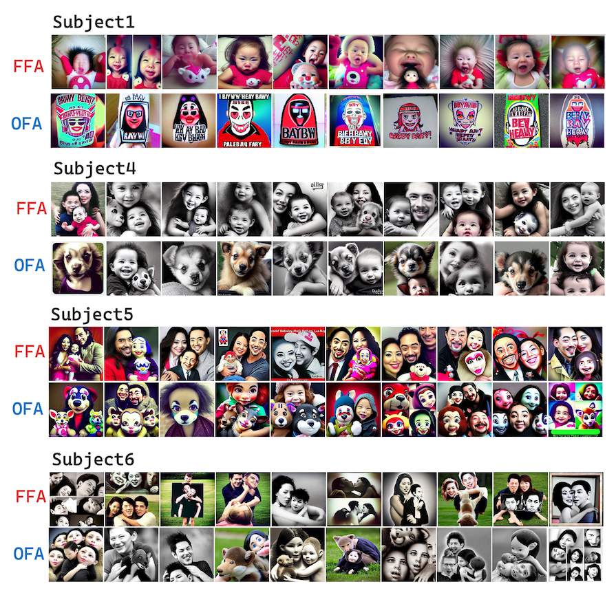
The reported results are predictions from these encoding models. In an evaluation using a different image encoder, the generated pictures had higher predicted activation than natural images. The outputs also differed between people. For one subject, images aimed at a face-sensitive area tended toward baby faces; for another, they tended toward groups of people. That variation is part of the point: the system searches for what a particular person’s model predicts, rather than assuming every brain uses visual information in exactly the same way.


If the pictures work in real brains
A vision-imaging clinic could one day use a person’s earlier scan to create a short sequence of tailored scenes. A face might become a group portrait, or a room might become a landscape, while researchers test a particular predicted pattern instead of cycling through a generic catalog of pictures. For scientists, that would replace much of the guesswork in choosing stimuli with targeted tests of how regions interact and how visual processing differs between people.
But that future depends on a test NeuroVolve has not yet passed. People must actually view the generated images in new fMRI experiments, and their measured responses must match the intended changes. The encoding models also have to remain accurate beyond the images used to train them. Personalized neural data would need safeguards as well. Until then, NeuroVolve has made multi-region brain objectives programmable in a model—not yet programmable in a living brain.

Sources
arXiv, “NeuroVolve: Evolving Visual Stimuli toward Programmable Neural Objectives”